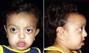
 クルーゾン病において視力障害や失明が発症する要因について、説明いたします。
クルーゾン病における失明の主な要因:
- 頭蓋内圧(intracranial pressure)の上昇 (ICP)による圧迫性の視神経萎縮:
- クルーゾン症候群では、頭蓋縫合の早期癒合(頭蓋骨癒合症)により頭蓋内圧が上昇することがあります。この圧力が直接に視神経を圧迫し、視神経萎縮を引き起こし、それが原因となって視力の低下や失明に繋がる可能性があります。また、頭蓋骨の異常な骨成長により、特に眼窩の後方や視神経管周辺で視神経が圧迫されうっ血乳頭なしに視神経障害(視神経萎縮)を引き起こして、視力低下が進行し、最終的には失明に至ることがあります。
- 鬱血乳頭 papilledema:
- 頭蓋内圧の上昇に伴い、クルーゾン症候群の患者は視神経軸索流にうっ滞を起こし鬱血乳頭(視神経乳頭の腫れpapilledema)を発症することがあります、鬱血乳頭ではその終末期に萎縮期を迎え、これが視神萎縮となって視力低下に寄与することがあります。
- 眼球突出 (眼球突出症):
- クルーゾン症候群では浅い眼窩(眼球の入る部分)が原因で眼球が突出(眼球突出)することがあります。この結果、眼球が外傷や乾燥にさらされ、角膜損傷が生じ、視力低下の要因となる可能性があります。
- 斜視と屈折異常:
- クルーゾン症候群では斜視(眼の位置のずれ)や顕著な屈折異常が見られることがあり、これが矯正されないと弱視(片眼の視力低下)を引き起こし、視覚障害の一因となる可能性があります。
- 網膜の変化:
- 頻度は少ないですが、上記の要因により網膜に変化が生じる場合もあり、それが失明に繋がることがあります。
対策:
- 早期介入: 頭蓋内圧の上昇の兆候を早期に監視し、頭蓋骨癒合症を修正する外科的介入を行うことで、視神経の損傷リスクを軽減できるという記載があります。
- 定期的な眼科評価: 定期的な眼科検査、特に視神経の画像診断が、進行性の変化を監視し管理するために不可欠です。
- 外科的治療: 重度の眼球突出や視神経圧迫がある場合、視力を維持するために眼窩減圧術などの外科的治療が必要となることがあります。
- 視覚リハビリテーション: 既に視力が低下している患者には、視覚リハビリテーションのサービスが残存視力を最大限に活用するために有益です。
これらのメカニズムを理解し、早期に介入することで、クルーゾン症候群の患者における失明リスクを軽減することが可能です。
以下に、クルーゾン病における失明や視覚障害のメカニズムについての理解に役立つ参考文献をいくつか挙げます。
- Crouzon Syndrome Overview:
- National Center for Biotechnology Information (NCBI) – PubMed Health:
- This source provides a comprehensive overview of Crouzon syndrome, including its genetic basis, clinical manifestations, and associated complications, including those related to vision.
- PubMed Health – Crouzon Syndrome
- National Center for Biotechnology Information (NCBI) – PubMed Health:
- Craniosynostosis and Vision:
- Mathijssen, I. M. J. (2015). “Guideline on Treatment and Management of Craniosynostosis.” Journal of Craniofacial Surgery, 26(6), 1735–1806.
- This article provides guidelines on the treatment of craniosynostosis, including the implications for vision and management of increased intracranial pressure.
- Link to Article
- Mathijssen, I. M. J. (2015). “Guideline on Treatment and Management of Craniosynostosis.” Journal of Craniofacial Surgery, 26(6), 1735–1806.
- Optic Neuropathy in Craniosynostosis:
- Thompson, D. N., Harkness, W., & Hayward, R. D. (1995). “Optic Nerve Compression in Craniosynostosis.” Pediatric Neurosurgery, 23(3), 155–160.
- This paper discusses the occurrence of optic nerve compression in craniosynostosis, including Crouzon syndrome, and its management.
- Link to Article
- Thompson, D. N., Harkness, W., & Hayward, R. D. (1995). “Optic Nerve Compression in Craniosynostosis.” Pediatric Neurosurgery, 23(3), 155–160.
- Ocular Findings in Crouzon Syndrome:
- Ragge, N. K., Subak-Sharpe, I. D., & Collin, J. R. O. (2007). “A Clinical, Molecular and Genomic Approach to Optic Atrophy in Craniosynostosis Syndromes.” Eye, 21(10), 1313–1320.
- This review article discusses the ocular findings in patients with craniosynostosis syndromes, including Crouzon syndrome, and the mechanisms leading to optic atrophy.
- Link to Article
- Ragge, N. K., Subak-Sharpe, I. D., & Collin, J. R. O. (2007). “A Clinical, Molecular and Genomic Approach to Optic Atrophy in Craniosynostosis Syndromes.” Eye, 21(10), 1313–1320.
- Increased Intracranial Pressure in Craniosynostosis:
- Fearon, J. A. (2009). “Evidence-Based Medicine: Craniosynostosis.” Plastic and Reconstructive Surgery, 124(6), 2061–2067.
- This article explores the relationship between increased intracranial pressure and craniosynostosis, including its impact on vision and the need for surgical intervention.
- 清澤注 ;対策として
-
- Fearon, J. A. (2009). “Evidence-Based Medicine: Craniosynostosis.” Plastic and Reconstructive Surgery, 124(6), 2061–2067.


コメント